Autologous Cultured Corneal Epithelium
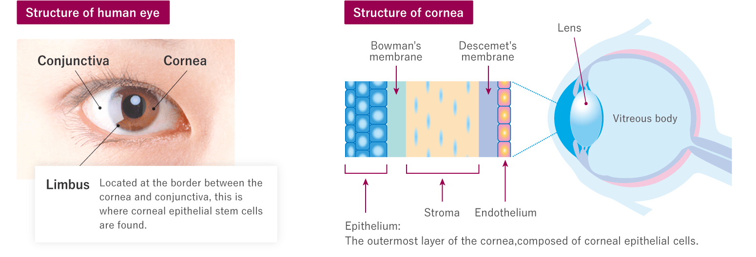
Corneal epithelium forms the outermost layer of the cornea in the eye. Within the corneal epithelium, stem cells are found in the region called the limbus on the border between the cornea and the conjunctiva. In cases of severe injury to the cornea, as long as even a small patch of healthy limbus remains, corneal epithelial stem cells can be isolated from this limbal tissue and cultured to produce autologous cultured corneal epithelium. Transplanting this tissue makes it possible for patients to recover their vision unheard of with conventional treatments.
Cultured Corneal Epithelium
Development
When the limbus is damaged the conjunctiva invade the cornea, resulting in scarring (conjunctivalization) of the cornea. Even transplantation of a cornea from a deceased donor has not proved a successful means of treatment, as the absence of limbal stem cells results in the worsening of symptoms. In 1997, Italian researchers Dr. Graziella Pellegrini and Dr. Michele De Luca demonstrated for the first time that transplanting cultured corneal epithelium into such patients suppressed the invasion of the conjunctiva to treat this condition.1) J-TEC has introduced the techniques developed by Dr. Pellegrini and her team for the development of cultured corneal epithelium.
1) Pellegrini G. et al., Lancet, 349, 990-993(1997).
Culture
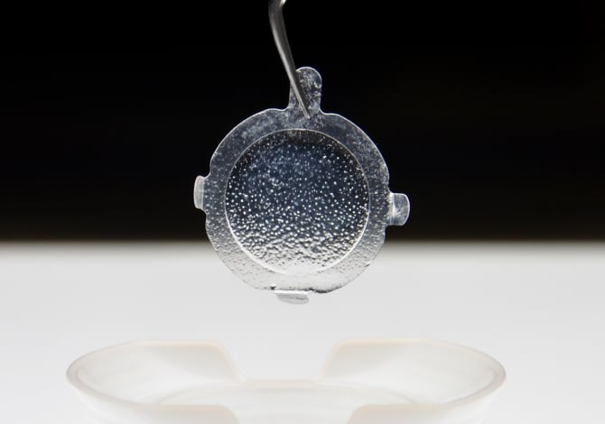
A 1-mm2 sample of the patient's own limbal tissue is taken, and cultured by the same method used to produce cultured epidermis. In other words, cells are enzymatically isolated from limbal tissue, and the isolated cells are cultured together with mouse fibroblasts in the same way as for cultured epidermis. In the final stage, corneal epithelial cells are cultured on a gel to produce the finished cultured corneal epithelium.
The use and commercialization of the medicinal products developed by Japan Tissue Engineering Co., Ltd. that are referred to on this website are approved only in Japan. A potential use and commercialization in other regions will be subject to the prior granting of a marketing authorization in the given territory and compliance with applicable laws.
Researchers
- Graziella Pellegrini, Ph.D.